MitraClip
MitraClip
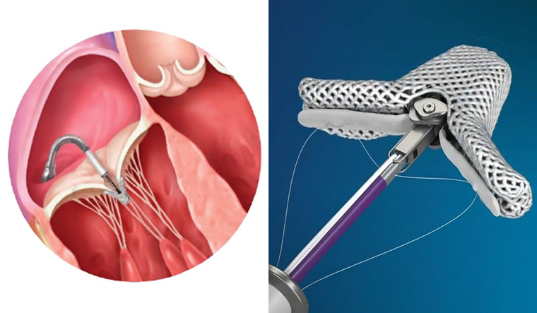
MitraClip is a breakthrough innovation for mitral valve patients. It is a device used to treat mitral valve regurgitation. This minimally invasive, transcatheter approach enables faster recovery and leading a better quality life.

MitraClip is a device used to repair leaky mitral valve – a condition called mitral regurgitation. Mitraclip device is inserted and positioned at the leaking portion of the valve through transcatheter technique. Unlike surgery, the MitraClip procedure does not require opening the chest and temporarily stopping the heart. Instead, doctors access the mitral valve with a thin tube (called a catheter) that is guided through a vein in your leg to reach your heart. The procedure is less invasive than traditional open-heart surgery and patients are usually released from the hospital within 2 to 3 days. Patients are known to experience improvement in their symptoms of mitral regurgitation and quality of life soon after the procedure.
MitraClip
In mitral regurgitation (MR) blood leaks backward to the heart caused by failure of the heart’s mitral valve leaflets to close tightly. This means that less blood is pumped out of the heart to supply the body. If the amount of MR is small and does not progress, the backward leak has no significant consequences.If significant (moderate to severe) MR is present, the left ventricle must work harder to keep up with the body’s demands for oxygen rich blood. Over time, the heart muscle undergo a series of changes to maintain this increased demand, depending upon the amount of blood that is regurgitated and how the heart responds to the regurgitated blood.
- Shortness of breath
- Fatigue
- Swelling in the legs etc.
Mitral valve prolapse –mitral valve leaflet tissue is deformed and elongated so that the leaflets do not come together normally. Other types of heart disease – MR can develop as a result of other types of heart diseases, such as after a heart attack or other cause of heart muscle injury. Congenital heart abnormality.
The Mitraclip repair procedure involves the following steps:
1. Vein puncture and guide catheter insertion Prior to sterile draping, a plate and a lift are placed under and over the right lower extremity respectively. From the femoral venous (femoral vein ) puncture, a TEE guided trans-septal puncture of the septum is performed using a standard kit with the needle, a dilator and a sheath. After successful septal puncture, the sheath is parked in the left atrium. The femoral vein entry site is dilated with a dilator
2. Steering/positioning of the guide catheter and CDS The Steerable Guide Catheter-Dilator assembly is inserted over the guide wire under TEE guidance. The Steerable Guide Catheter handle is secured in the sterile stabilizer placed on top of the previously placed lift. The dilator and guidewire are removed together and the guide catheter is de-aired. The Clip Delivery System (CDS) is inserted through the Steerable Guide Catheter under fluoroscopic and TEE guidance The Steerable Guide Catheter and Clip Delivery System (CDS) are positioned in the left atrium for clip deployment using echocardiographic and fluoroscopic guidance. The clip arms are opened and the clip is positioned The Steerable Guide Catheter-Dilator assembly is inserted over the guide wire under TEE guidance. The Steerable Guide Catheter handle is secured in the sterile stabilizer placed on top of the previously placed lift
3. Leaflet grasping, leaflet insertion assessment, and clip closure After aligning the CDS, and the MitraClip, the mitral valve leaflets are grasped and the MitraClip is partially closed to reduce MR Quality of the grasp, valve function and adequacy of repair (reduction of valve leak) are assessed using echocardiography and fluoroscopy if desired.
4. MitraClip deployment and system removal. The clip is closed further as needed under real time MR assessment. If necessary, the Clip is repositioned to reduce MR further A second clip may be placed as needed to further reduce MR
5. The steerable guide catheter is removed and groin access is closed.
Anatomic suitability is assessed by transesophageal echocardiography. Factors such as the origin of mitral regurgitation, the flail segment width are taken into consideration.
- Quicker recovery, Less pain and Less discomfort
- Minimally invasive as compared to open-heart surgery
- Average stay of patient is 2 – 3 days, however it may vary depending on the condition of patient
- Improvement in the symptoms (shortness of breath and low pressure etc.) of mitral regurgitation can be felt immediately after the procedure
- Improved lifestyle of the patient
Book Appointment


Our cardiologist provides top-notch care tailored to your individual needs. From prevention to treatment, we ensure your heart is in the best hands.
About Doctor
For Appointment
Contact us
