Ventricular Septal Defect (VSD)
Ventricular Septal Defect (VSD)
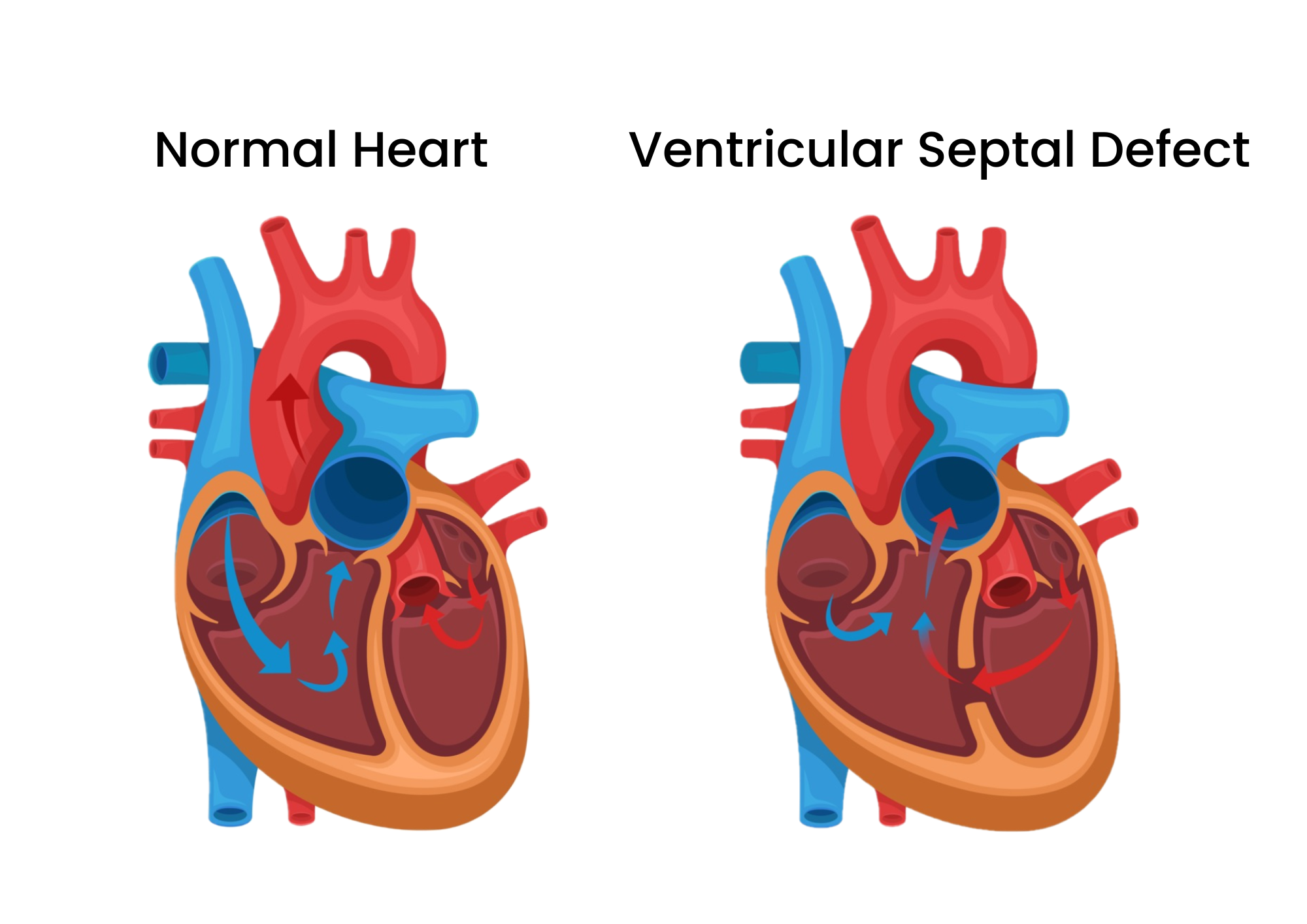
A ventricular septal defect (VSD) is a hole in the heart that's present at birth (congenital heart defect). The hole is between the lower heart chambers (right and left ventricles). It allows oxygen-rich blood to move back into the lungs instead of being pumped to the rest of the body.

A ventricular septal defect (VSD) changes how blood flows through the heart and lungs. Oxygen-rich blood gets pumped back to the lungs instead of out to the body. The oxygen-rich blood mixes with oxygen-poor blood. These changes may increase blood pressure in the lungs and require the heart to work harder to pump blood.
A small ventricular septal defect may cause no problems. Many small ventricular septal defects (VSDs) close on their own. Babies with medium or larger VSDs may need surgery early in life to prevent complications
Ventricular Septal Defect (VSD)
Ventricular Septal Defect (VSD) is a congenital heart defect characterized by an abnormal opening in the wall (septum) that separates the two lower chambers (ventricles) of the heart. This defect allows oxygen-rich blood from the left ventricle to mix with oxygen-poor blood from the right ventricle. This can lead to increased workload on the heart and increased blood flow to the lungs, potentially causing complications over time.
- Perimembranous VSD: Located in the upper part of the ventricular septum, near the heart valves. This is the most common type.
- Muscular VSD: Found in the lower part of the septum, which is the muscular part. These defects can be multiple and are often referred to as “Swiss cheese” VSDs.
- Inlet VSD: Located close to where the blood enters the ventricles, near the atrioventricular valves.
- Outlet (or Conal) VSD: Located in the outflow tract of the ventricles, near where the blood exits to the major arteries.
- Shortness of breath
- Rapid breathing or breathlessness
- Fatigue
- Poor growth in infants
- Sweating during feeding or playing
- Frequent respiratory infections
VSD is often diagnosed through:
- Echocardiogram: An ultrasound of the heart, which is the primary tool for diagnosing VSD.
- Electrocardiogram (ECG): Measures the electrical activity of the heart.
- Chest X-ray: Provides images of the heart and lungs.
- Cardiac MRI: Detailed images of the heart.
- Cardiac catheterization: Invasive test to measure heart pressures and oxygen levels.
- Monitoring: Small VSDs may close on their own or not cause significant problems.
- Medications: To manage symptoms such as heart failure or high blood pressure in the lungs.
- Surgery: To close the defect, often recommended for larger VSDs or if symptoms are present. Surgery may involve stitching the hole or placing a patch over it.
- Catheter Procedures: For some types of VSDs, a device can be inserted via a catheter to close the defect without open-heart surgery.
Book Appointment


Our cardiologist provides top-notch care tailored to your individual needs. From prevention to treatment, we ensure your heart is in the best hands.
