Atrial Septal Defect (ASD)
Atrial Septal Defect (ASD)
The normal human heart is responsible for efficiently receiving and pumping blood. Proper blood flow is vital to health because the body's tissues rely on the blood to carry nourishment (oxygen, glucose) and remove waste products (carbon dioxide). Under normal conditions, the cardiovascular system (heart, blood vessels) contains two separate circulatory systems - venous (right) and arterial (left). Venous (deoxygenated, blue) blood returns from the body to the right atrium.

Blood flows from the right atrium through the tricuspid valve into the right ventricle where blood is actively pumped across the pulmonary valve and into the pulmonary arteries. The main pulmonary artery divides into large right and left pulmonary arteries and then to small pulmonary arterioles. Adjacent to the small arterioles, numerous pulmonary air filled sacs. Within these small sacs, gases are exchanged. Waste products from the venous blood enter the air sacs and are removed from the pulmonary system when we breathe out (exhale). Oxygen that enters the lungs when we breathe in (inspiration) enters the small air sacs and diffuses into the blood. Oxygen-rich (red) blood returns from the lungs via pulmonary veins to the left atrium.
Muscular and connective tissues separate the right (blue blood) from the left (red blood) side of the heart. The heart is separated at the level of the receiving chambers (right and left atria) by the atrial septum, and at the pumping chambers (right and left ventricles) by the ventricular septum. Persistent opening between the atrial and ventricular septa in postnatal life is abnormal.
Atrial Septal Defect (ASD)
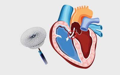
Atrial Septal Defect (ASD) is a congenital heart defect characterized by a hole in the wall (septum) that separates the two upper chambers (atria) of the heart. This opening allows oxygen-rich blood to flow from the left atrium into the right atrium, mixing with oxygen-poor blood. As a result, the heart and lungs have to work harder, which can lead to complications over time.
- Secundum ASD: The most common type, located in the middle of the atrial septum.
- Primum ASD: Located in the lower part of the atrial septum, often associated with other heart defects.
- Sinus Venosus ASD: Located near the junction of the right atrium and the superior or inferior vena cava.
- Coronary Sinus ASD: A rare type, located within the coronary sinus, a collection of veins that join together to form a large vessel that collects blood from the heart muscle.
- Shortness of breath
- Fatigue
- Swelling of legs, feet, or abdomen
- Heart palpitations or skipped beats
- Stroke
ASD is often diagnosed through:
- Echocardiogram: An ultrasound of the heart.
- Electrocardiogram (ECG): Measures the electrical activity of the heart.
- Chest X-ray: Provides images of the heart and lungs.
- Cardiac MRI: Detailed images of the heart.
- Cardiac catheterization: Invasive test to measure heart pressures and oxygen levels.
- Monitoring: Small ASDs may close on their own or not cause significant problems.
- Medications: To manage symptoms such as irregular heartbeats.
- Surgery or Catheter Procedures: To close the defect, often recommended for larger ASDs or if symptoms are present.
Book Appointment


Our cardiologist provides top-notch care tailored to your individual needs. From prevention to treatment, we ensure your heart is in the best hands.
About Doctor
For Appointment
Contact us
